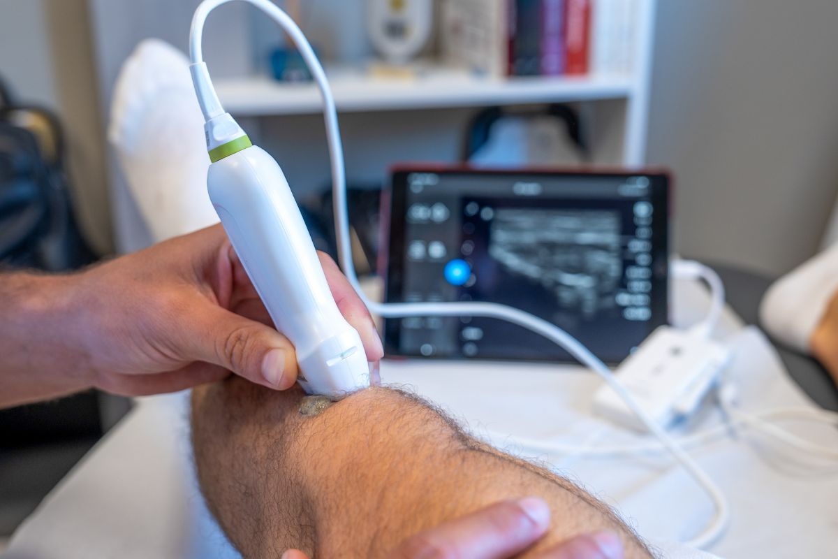
What is musculoskeletal ultrasound?
Musculoskeletal ultrasound is an imaging technique used to look at structures such as tendons, muscles, ligaments, bursae and some joint-related problems.
It uses sound waves to create live images of the area being assessed. There is no radiation involved, and the scan can usually be performed during a clinic appointment.
Ultrasound can be particularly helpful when assessing soft tissue pain, tendon irritation, swelling, bursitis, fluid around a joint or certain sports-related injuries.
It may also be used to guide injections accurately to the intended area.
When might ultrasound be useful?
Ultrasound may be helpful if you have pain, swelling, stiffness or reduced function affecting a joint, tendon or soft tissue area.
It is commonly used in sports medicine and MSK care to assess problems such as tendon pain, tendon tears, bursitis, ligament sprains, muscle injuries, joint swelling and soft tissue irritation.
It can also be useful when symptoms are very specific to one area, or when examination findings need to be supported by imaging.
For some patients, ultrasound can help confirm the diagnosis. For others, it may help decide whether further imaging, treatment, rehabilitation or referral is needed.
What can ultrasound assess?
Musculoskeletal ultrasound may be used to assess a range of areas and conditions, including:
Tendon pain and tendinopathy
Tendon tears or thickening
Bursitis and soft tissue inflammation
Muscle injuries
Ligament sprains
Joint swelling or fluid
Soft tissue lumps or localised swelling
Guidance for injections
Some causes of shoulder, elbow, wrist, hip, knee, ankle and foot pain
The exact use of ultrasound will depend on your symptoms, the area involved and what Dr Jones is looking to understand from the assessment.
Ultrasound and guided injections
Ultrasound can also be used to guide injection treatments.
This allows the needle position to be viewed during the procedure, helping the injection reach the intended joint, tendon sheath, bursa or soft tissue target.
Image guidance can be particularly useful where accuracy matters, where the anatomy is more complex, or where previous treatment has not helped as expected.
Depending on the diagnosis, ultrasound-guided injections may include corticosteroid, hyaluronic acid or platelet-rich plasma treatment.
Ultrasound compared with MRI or X-ray
Ultrasound is not always the right scan for every problem.
X-rays are often useful for looking at bones, joint space and arthritis. MRI can give a more detailed view of deeper structures inside a joint or around the spine.
Ultrasound is particularly useful for many soft tissue problems because it is quick, dynamic and can be used during the appointment.
If another scan is more appropriate, this can be discussed as part of your assessment.
What happens during an ultrasound assessment?
During the scan, gel is applied to the skin and a handheld ultrasound probe is moved over the area being assessed.
You may be asked to move the joint or muscle during the scan, or to point to the exact area of pain.
The scan is usually comfortable and does not involve radiation.
Findings can often be discussed during the appointment, alongside your symptoms, examination and treatment options.
After your ultrasound
After your ultrasound assessment, Dr Jones can explain what the scan shows and how it fits with your symptoms and examination.
In some cases, the scan may help confirm a diagnosis and guide a treatment plan. In others, it may show that further imaging or a different specialist opinion is needed.
The aim is to use ultrasound as part of a wider clinical assessment, helping you understand the problem and make a clear decision about the next step.
Book an ultrasound assessment
If you are struggling with joint pain, tendon symptoms, soft tissue pain or a sports-related injury, ultrasound assessment may help clarify the diagnosis and guide treatment.
Dr Jones can assess your symptoms, discuss whether ultrasound is appropriate and advise on the most suitable next step.