Detailed imaging when a clearer view is needed
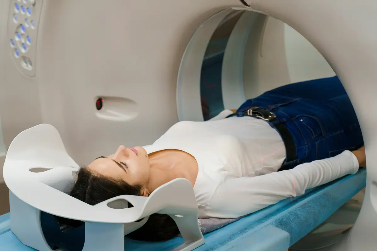

A CT scan is a type of imaging used to create detailed cross-sectional pictures of the body.
In musculoskeletal and sports medicine, CT can be useful when more detail is needed than a standard X-ray can provide, particularly for bones, joints, fractures, alignment and more complex structural problems.
Dr Jones may recommend a CT scan when symptoms, examination findings or previous imaging suggest that a more detailed view would help guide the next step.
The aim is to use CT imaging appropriately, alongside your history, examination and goals, so that treatment decisions are based on a clearer understanding of the problem.
A CT scan uses X-rays and computer technology to create detailed images of the body in thin slices.
These images can be viewed from different angles and can give a much clearer picture of bone and joint structure than a standard X-ray.
CT scans are particularly helpful when looking at complex fractures, subtle bone changes, joint surfaces, alignment, previous surgery or areas where a more detailed view is needed.
CT is not always the first scan used, but it can be very valuable when the diagnosis is unclear or when treatment planning needs more detail.
A CT scan may be helpful if you have persistent joint pain, suspected bone injury, symptoms after trauma, or a problem that has not been fully explained by an X-ray or examination.
It may also be used when a fracture is difficult to see clearly, when joint surface detail matters, or when more information is needed before referral to another specialist.
CT can be particularly useful for assessing areas such as the ankle, foot, hip, knee, shoulder, wrist and spine, depending on the symptoms and clinical question.
The decision to arrange a CT scan is based on whether the extra detail is likely to change the diagnosis, treatment plan or referral pathway.
CT imaging can help assess a range of bone and joint problems, including:
The findings are always considered alongside your symptoms and physical assessment.
A scan can show structural changes, but the most useful treatment plan comes from matching the imaging with how the problem is affecting you.
CT scans can be helpful when joint pain is linked to structural change, previous injury, arthritis, bone shape or alignment.
They may be used when X-rays show a problem that needs closer assessment, or when symptoms suggest something more detailed may be happening within the joint.
In some cases, CT can help show the shape of the joint surface, bony changes, loose fragments, or how a previous injury has healed.
This can be useful when deciding whether physiotherapy, injection treatment, bracing, further imaging or surgical referral may be appropriate.
Different scans are useful for different reasons.
X-rays are often the first step for looking at bones, joints, arthritis and alignment.
Ultrasound is useful for many tendon, muscle, ligament, bursa and soft tissue problems, and can also be used to guide injections.
MRI gives more detail about soft tissues, cartilage, bone marrow, discs, ligaments and structures inside a joint.
CT is particularly useful when detailed bone and joint structure needs to be assessed in more depth than a standard X-ray can provide.
Dr Jones can advise which scan is most suitable based on your symptoms, examination and what needs to be understood.
A CT scan is usually quick and straightforward.
You will normally lie on a scanning table, which moves slowly through the scanner while images are taken.
The scan itself is usually painless and often only takes a few minutes, although the full appointment may take longer.
In some cases, contrast dye may be used to highlight certain structures, but this depends on the reason for the scan and will be explained before the appointment if required.
CT scans use radiation, so they are only recommended when the expected benefit outweighs the exposure involved.
For many musculoskeletal problems, the amount of radiation is carefully controlled and the scan is targeted to the area being assessed.
Dr Jones will only recommend CT imaging where it is likely to provide useful information for diagnosis, treatment planning or referral.
If another type of imaging would be more appropriate, this can be discussed.
After your CT scan, the images and report can be reviewed alongside your symptoms, examination findings and any previous imaging.
Dr Jones can explain what the scan shows, whether the findings match your symptoms and what the next step may be.
This may include advice, physiotherapy, activity modification, injection treatment, further imaging or referral to an orthopaedic surgeon or another specialist where needed.
The aim is to turn the scan result into a clear, practical plan.
If you are struggling with joint pain, injury-related symptoms, persistent pain after trauma or an MSK problem that needs further investigation, an assessment can help decide whether CT imaging is appropriate.
Dr Jones can review your symptoms, arrange suitable imaging where needed and guide the next step in your treatment plan.
We use cookies and similar technologies to improve your experience on our website. Read our Privacy Policy.